£1,010.00 ex. Vat
Available on backorder
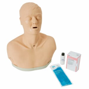
Paramedics, EMTs, combat medics, flight nurses, anaesthesiologists and other emergency medical personnel will have the opportunity to strengthen their ability and confidence to perform or assist in implementing surgical airways. Anatomically accurate landmarks aid in site training and allow for fast action.
The hyperextended neck allows the user to determine the proper incision site. The trachea in the simulator is replaceable, as the airway passes completely through from top to bottom. This allows checking the stylet and obturator placement once the incision has been made.
Complete with a chin and full-size neck, use ties to hold the obturator in a secure position. Inflation of the simulated lung verifies correct placement.
A cricothyrotomy is often used as an airway of last resort given the numerous other airway options available including standard tracheal intubation and rapid sequence induction, which are the common means of establishing an airway in an emergency scenario. Cricothyrotomies account for approximately 1% of all emergency department intubations and are used mostly in patients who have experienced a traumatic injury.
Some general indications for this procedure include:
The procedure was first described in 1805 by Félix Vicq-d’Azyr, a French surgeon and anatomist. A cricothyrotomy is generally performed by making a vertical incision on the skin of the throat just below the laryngeal prominence (Adam’s apple), then making another transverse incision in the cricothyroid membrane which lies deep to this point. A tracheostomy tube or endotracheal tube with a 6 or 7-mm internal diameter is then inserted, the cuff is inflated, and the tube is secured. The person performing the procedure might utilise a bougie device, a semi-rigid, straight piece of plastic with a one-inch tip at a 30-degree angle, to provide rigidity to the tube and assist with guiding its placement.
Confirmation of placement is assessed by bilateral auscultation of the lungs and observation of the rise and fall of the chest. Alternatively, bedside ultrasound has been used increasingly to guide the procedure and confirm the placement of the tracheal tube. It may especially be helpful in situations where a neck collar is placed.
This procedure is rarely performed, given advancements in airway technique and adjuncts, and thus simulated training is of paramount importance to correctly perform this procedure under a high-stress situation.


Simulaids is a trusted supplier to the following brands:


Sign up to our newsletter
Sign up to our newsletter for the latest product updates, announcements, and insights.
© Simulaids 2024. All Rights Reserved.